

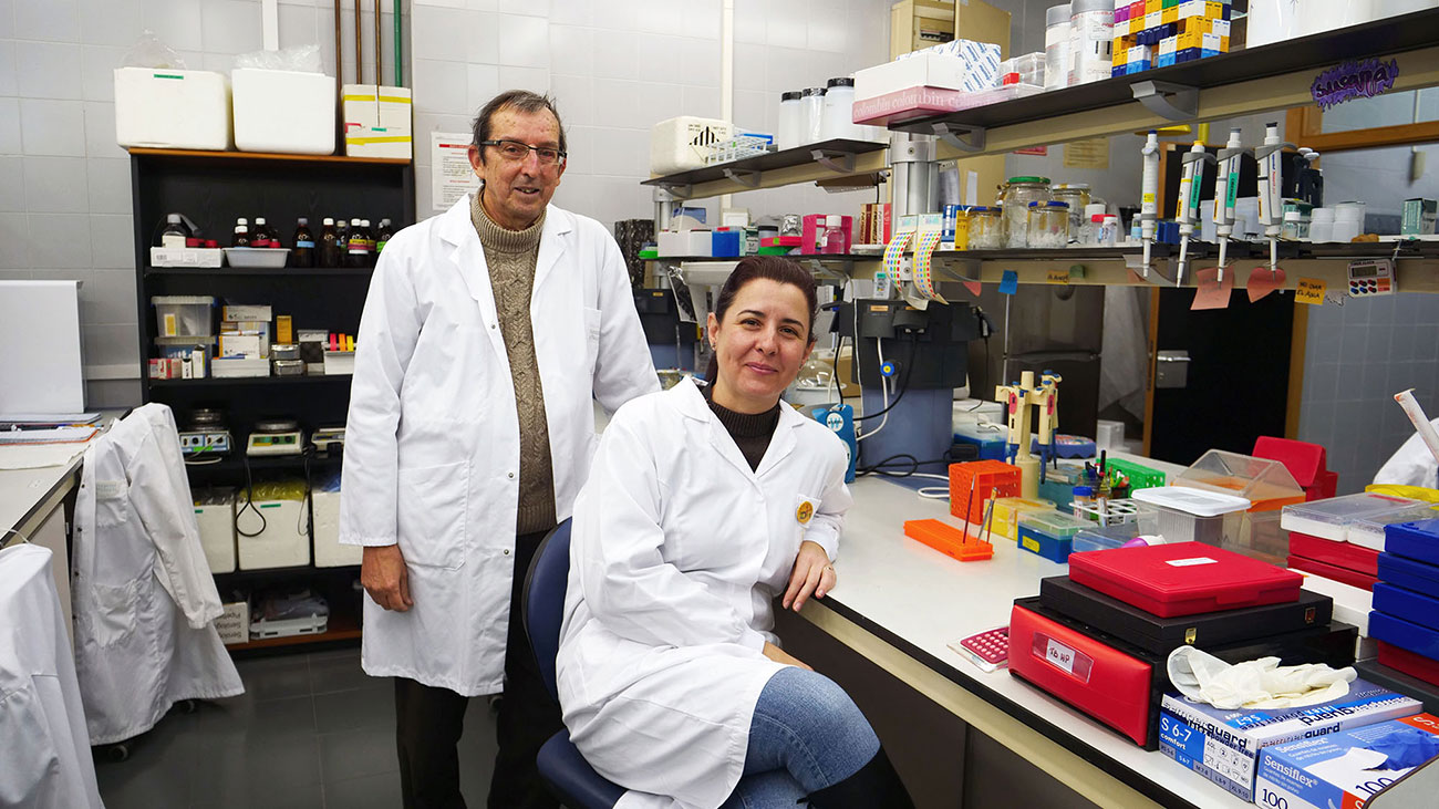
J.M. García-Verdugo and Susana González
An international research team has identified, in the brain of foetuses, a population of neuroblasts that migrate to the cerebral cortex and generate inhibitory-type interneurons. This means that they have discovered the starting point of a cellular organisation that is essential for the proper functioning of the brain. Led by the University of San Francisco, the work has the participation of the Cavanilles Institute of the University of Valencia-CIBERNED, as well as the Health Research Institute (IIS) and the La Fe University and Polytechnic Hospital in Valencia.
The cerebral cortex is the most complex region of the human brain. Emotions, speech, thought itself emanate from it. Its functions are regulated by the balance between the activity of the excitatory neurons, which are activated to send signals to other cells, and the inhibitory ones, which fine-tune the activity of the former. Both are essential to maintain brain balance.
To understand the functioning of the cerebral cortex, in normal and pathological conditions, it is necessary to know where its embryonic cells are generated, how they are organised or how their migration occurs from the place of origin to the cerebral cortex. This route is essential, since if they do not reach their destination or migration defects occur, the brain is destabilised and pathologies such as autism, epilepsy or schizophrenia may appear. They are neurodevelopmental disorders that affect more than 5% of the population.
The work that Science has just published and that the magazine has described as a highlighting or relevant article accurately identifies, in the prenatal brain, a population of neuroblasts – embryonic precursor cells of neurons – that migrate to the cerebral cortex and generate inhibitory-type interneurons.
These neuroblasts – here the main finding of the scientific team – are located in the so-called Medial Ganglionic Eminence (MGE), an anatomical prominence that contacts the ventricular cavity of the human brain. “Although this migration was known in mice, it was not known in detail how they were organised in this region in humans”, comments José Manuel García Verdugo, Professor of Cell Biology at the University of Valencia and one of the senior researchers in the project. García Verdugo is also responsible, together with the biologist Susana González (CIBERNED), for the electronic microscopy of the research. “We are now studying the details of this migration and, although the results of this work will not have short-term application for the cure of pathologies related to inhibitory neurons, a deeper understanding of this process will help to understand the behaviour of neuroblasts during development, as well as its implication in associated pathologies”, concludes the scientist.
The study
Entitled “Nests of dividing neuroblasts sustain interneuron production for the developing human brain”, the article in Science explains that the scientific team sought to characterise the organisation of these cells in the EGM, observing the formation of small groups of highly proliferating cells associated with each other. Between these groups, as the text indicates, expansions of radial cells are interspersed in the form of columns that start from the ventricular surface and that mark, as guides, the path of migration towards the cerebral cortex.
In addition, the researchers transplanted these cell populations into the cerebral cortex of young mice, observing that the cells migrate through cortical and subcortical regions while continuing to divide and differentiate into mature interneurons. “Synchronising the obtaining of cells for transplantation and conditioning young mice as recipients has been a challenge for clinical and research teams”, explains Máximo Vento, head of the Perinatology Research Group at IIS La Fe. His team and the Obstetrics and Pathological Anatomy services of Hospital La Fe have been in charge of identifying the cases, obtaining and processing, and supplying the samples to the scientific team for the study. “Another challenge has been obtaining post-mortem samples in optimal condition”, adds Jaime Ferrer, a specialist in Anatomical Pathology. “These are samples from fetuses incompatible with life, provided thanks to the generosity of the affected families”, he concludes.
Reference:
Nests of dividing neuroblasts sustain interneuron production for the developing human brain. Mercedes F. Paredes, Cristina Mora, Quetzal Flores-Ramírez, Arantxa Cebrián-Silla, Ashley Del Dosso, Phil Larimer, Jiapei Chen, Gugene Kang, Susana González Granero, Eric García, Julia Chu, Ryan Delgado, Jennifer A. Cotter, Vivian Tang, Julien Spatazza, Kirsten Obernier, Jaime Ferrer Lozano, Máximo Vento, Julia Scott, Colin Studholme, Tomasz J Nowakowski, Arnold R. Kriegstein, Michael C. Oldham, Andrea Hasenstaub, Jose Manuel Garcia-Verdugo, Arturo Alvarez-Buylla, Eric J. Huang.
https://doi.org/10.1126/science.abk2346