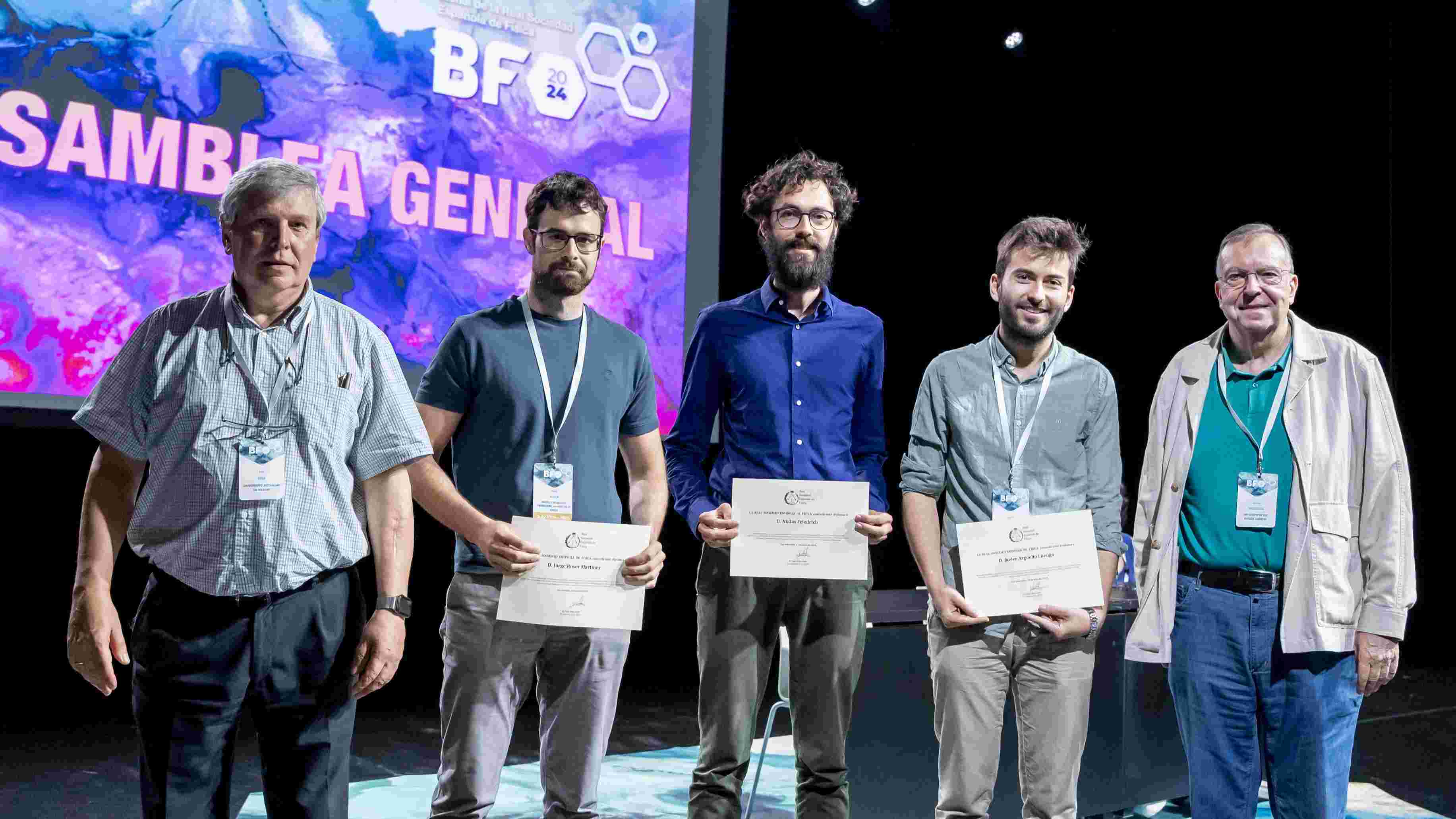
Jorge Roser's doctoral thesis, carried out at the Institute of Corpuscular Physics (IFIC), has been awarded the prize of the Spanish Royal Society of Physics (RSEF) for the best doctoral thesis in the Applied Physics category. Her work, entitled “Improving Image Quality in a multi-plane Compton Telescope for Hadron Therapy Monitoring”, had already been previously awarded by the Medical Physics Specialized Group of the RSEF, which allowed her to opt for the general awards of the institution
The award ceremony took place during the celebration of the XXXIX Biennial Meeting of the RSEF, hosted in the city of San Sebastian from 15 to 19 July 2024. Roser received the award from the hands of the President of the RSEF, Luis Viña Liste.
Roser's thesis, directed initially by Josep Oliver and in recent years by Gabriela Llosá, falls within the field of hadronic therapy, a new type of cancer therapy that, unlike conventional X-ray radiotherapy, uses heavy charged particles, generally protons (proton therapy) or, to a lesser extent, carbon ions. This research explores various methods to improve the quality of images obtained with Compton cameras, with the aim of applying them to real-time range finding in hadronic therapy.
This application presents significant challenges, as it requires detecting relatively high energy gamma rays emitted over a wide energy range along with other unwanted particles. In addition, the Compton camera must withstand very high emission rates during short detection times. In return for such difficulties, the potential advantages of real-time range-checking are promising, as it would allow exploiting the dosimetric benefits of hadron therapy over conventional radiotherapy, thus improving the quality of life of patients treated with this type of therapy.
Roser's work focuses on the experimental evaluation of analytical models, retrieval strategies and modifications in reconstruction algorithms with MACACO, a new Compton camera prototype developed by the IRIS group at IFIC, located at the University of Valencia Science Park. A crucial aspect of their research is the full use of the capabilities of this prototype to obtain the best possible image quality. In this sense, a model of the System Matrix and sensitivity has been developed to obtain the image with three-interaction events, and modifications have been implemented on the existing models of the group to integrate the different information channels available in MACACO or to improve its accuracy. In addition, a modification of the image reconstruction algorithm has been proposed to integrate these information channels into a single reconstruction process. As a result, a remarkable improvement in the quality and robustness of the obtained images has been found.
With all the proposed improvements, the MACACO prototype has proven to be able to experimentally detect displacements in the range of up to 2 mm in dummies irradiated with proton beams for clinical energies, which is a significant improvement over the results obtained to date.
Jorge Roser's scientific career
Roser began her academic career in 2012, when she started the Degree in Physics at the University of Valencia, completing it in 2016. In 2017, she obtained a Master's degree in Advanced Physics at the same university, at which time she developed a deep interest in medical physics. This led him to start a PhD with the IRIS group at IFIC, focused on image reconstruction with Compton cameras for range-finding in hadronic therapy. After finishing her PhD thesis in 2023, Roser is currently doing a postdoctoral stay at the Institute of Medical Engineering at the University of Lübeck in Germany.
IFIC IRIS Group
The IRIS group (Image Reconstruction, Instrumentation and Simulations for medical applications), coordinated by IFIC researcher Gabriela Llosá Llácer, specializes in the development of detectors for medical applications. The research team has focused its efforts on medical imaging and, in particular, on the monitoring of hadronic therapy or the verification of radiopharmaceutical treatments, in the latter case with the aim of improving the visualization of their distribution in the human body when administered to the patient.


