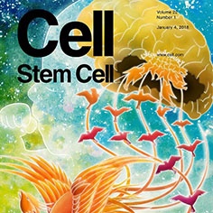
Researchers from the Universitat de València and the Nagoya University (Japan) have discovered a neural regeneration mechanism following brain injuries by means of rat testing. This mechanism only takes place during the neonatal period. This discovery, appearing on the front page of the magazine Cell Stem Cell, permits developing new therapies that can ease the consequences of ischemic events in the brain, which is one of the top challenges in Medicine nowadays.
The development of perinatal care in the past decades has enormously improved survival rate among new-borns. However, brain disorders such as hypoxic ischaemic encephalopathy (a disorder due to the lack of oxygen in the cerebral blood flood), which causes severe neural damage, still affect an important group of new-borns. Currently, a procedure to regenerate damaged neurones does not exist. Therefore, one of the top challenges of modern Medicine is developing new therapies that can ease the consequences of cerebral ischemic events.
José Manuel García-Verdugo, a researcher from the Cavanilles Institute of Biodiversity and Evolutionary Biology at the Universitat de València, located at the Scientific Park, and Kazunobu Sawamoto (Faculty of Medicine at the Nagoya University) have discovered a neural regeneration mechanism following brain injuries by means of rat testing. This mechanism only takes place during the neonatal period. “We have found out by means of rat testing that radial neuroglia that normally vanishes after the birth remains in neonatal cerebral injuries” indicates García Verdugo. Neurones produced via brain stem cells adhere to radial neuroglia through N-cadherin molecules by efficiently shifting along glia radial fibre towards the damaged area.
Cells that act like stem cells during the brain developing period, known as “radial glia”, have a unique molecular morphology with long radial fibres. Glia radial fibres act as a road for neuronal migration during the embryonic phase, and they disappear immediately after birth. Nonetheless, after a brain injury, the radial glia remains. This recent study has discovered that neurones produced from stem cells in the brain migrate efficiently to the damaged area through long radial glia fibres. In addition, by transplanting roads based on gelatine sponges that artificially simulate radial glia in the neonatal damaged brain, it is possible to foster neuronal movement towards the damaged area and restore the motion function of new-born mice. This neural regeneration mechanism is likely to be implemented in regenerative medicine for cerebral neonatal injuries in humans. “It is necessary to develop new therapies to ease cerebral neonatal disorders that can be used in the clinical practice in the future” adds García Verdugo.
Researcher Vicente Herranz-Pérez from Universitat de València has participated in this study that has been conducted by a partnership agreement between both universities and has been funded by the Japan Society for the Promotion of Science (JSPS) in order to improve exchanges among researchers in key areas at an international scale. This study has appeared on the front page of the magazine Cell Stem Cell.
Reference:
Radial Glial Fibres Promote Neuronal Migration and Functional Recovery after Neonatal Brain Injury. Hideo Jinnou, Masato Sawada, Koya Kawase, Naoko Kaneko, Vicente Herranz-Pérez,Takuya Miyamoto,Takumi Kawaue,Takaki Miyata,Yasuhiko Tabata,Toshihiro Akaike,José Manuel García-Verdugo,Itsuki Ajioka, Shinji Saitoh and Kazunobu Sawamoto.
The front page shows a brain from a new-born who suffered an injury due to the lack of oxygen during the birth. The phoenix in the lower left corner of the image symbolizes the regeneration source in the cerebral damaged area. Feathers located in the tail of the phoenix represent radial glia cells acting as a road that leads young neurones to the damaged area. They are illustrated as little birds flying towards the brain. Once they get to the damaged area, young neurones integrate and generate mature neurones.


