

A research group of the Instituto de Física Corpuscular (IFIC), a joint center of the Consejo Superior de Investigaciones Científicas (CSIC) and the Universitat de València, develops a photon detection and imaging system that will allow to visualize during a medical treatment the distribution of the radiopharmaceutical in the patient's body and, in this way, verify that the radiopharmaceutical accumulates in the expected place and better estimate the radiation dose received by the tumor and the other organs
The Image Reconstruction, Instrumentation and Simulations for medical applications (IRIS) group of the Instituto de Física Corpuscular (IFIC), belonging to the scientific-academic area of the Parc Científic de la Universitat de València, is coordinated by the institute's researcher Gabriela Llosá Llácer, and specializes in the development of detectors for medical applications. The research team focused its efforts on medical imaging and, specifically, on the monitoring of hadronic therapy or the verification of radiopharmaceutical treatments, in the latter case, with the aim of improving the visualization of their distribution in the human body when administered to the patient.
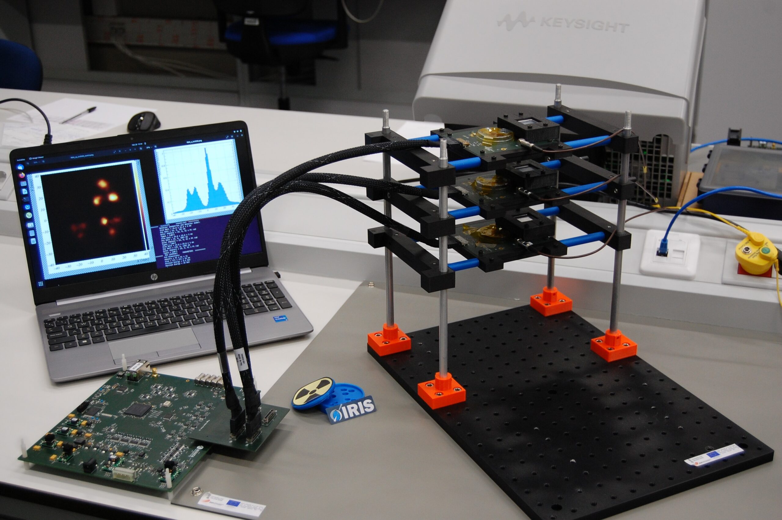
The technology developed by the IFIC team consists of a photon detection and imaging system based on lanthanum bromide crystals coupled to silicon photomultipliers that uses two or three detectors in time coincidence and, compared to conventional systems, offers higher detection efficiency, very good spatial resolution and a large field of view with a compact detector.
The technology developed by the IFIC team consists of a photon detection and imaging system based on lanthanum bromide crystals coupled to silicon photomultipliers using two or three detectors in time coincidence.
As Llosá explains, "the photon imaging systems currently in use generally have low efficiency, and have greater difficulty when there are photons of different energies, when these energies are higher than those of the radiotracers used for diagnosis, or, in certain areas, when the energy of the incident photons is unknown."
"We have devised and patented a noise reduction method that allows us to work in adverse low-signal scenarios, and we are also using artificial intelligence for image enhancement," adds Llosá.
For his part, Jorge Roser, CSIC researcher at IFIC, stresses that "to find out the incident energy of the photons in our detector, we have developed analytical models of image formation that improve the traditional reconstruction algorithms and allow us to obtain four-dimensional images, where the fourth dimension is the energy of the incident gamma rays."
"We have devised and patented a noise reduction method that allows us to work in adverse low-signal scenarios and we are also using artificial intelligence for image enhancement," Gabriela Llosá Llácer, IFIC researcher and project coordinator.
Previous applications
This technology has been previously used in astroparticle experiments or, for example, for the detection of radioactive foci after nuclear accidents on board drones or robots.
In the medical field, the IRIS group has carried out experiments in collaboration with proton therapy centers such as Quirónsalud (Madrid) for the monitoring of hadronic therapy, and with La Fe hospital (Valencia) for the verification of treatment with radiopharmaceuticals. At present, this technology has entered a valorization phase in which the device is being tested in relevant environments to increase its TRL (method for estimating the progress of a technology), and the aim is to arouse the interest of companies to ensure future commercialization of the device. This is carried out within the VALMONT (INNVA1/2021/37) and VALID (PDC2021-121839-I00) projects, funded by the Valencian Innovation Agency and the State Research Agency, respectively.